

Rigorous Methods for Stem Cell-based Models of Neuropathic Pain: Information For Researchers
Authors
- Pascal Röderer, PhD, Institute of Neurophysiology, Uniklinik RWTH Aachen, Aachen, Germany; Scientific Center for Neuropathic Pain Aachen SCNAACHEN, Uniklinik RWTH Aachen, Aachen, Germany.
- Hajira Elahi, PhD, Department of Pain Medicine, University of Texas MD Anderson Cancer Center, Houston, TX, USA.
- Patrick Dougherty, PhD, Department of Pain Medicine, University of Texas MD Anderson Cancer Center, Houston, TX, USA.
- Angelika Lampert, MD, Institute of Neurophysiology, Uniklinik RWTH Aachen, Aachen, Germany; Scientific Center for Neuropathic Pain Aachen SCNAACHEN, Uniklinik RWTH Aachen, Aachen, Germany.
- Franziska Denk, DPhil, Wolfson Sensory, Pain and Regeneration Centre (SPaRC), Guy’s Campus, King’s College London.
- Margarita Calvo, PhD, Faculty of Biological Sciences, Pontificia Universidad Católica de Chile, Santiago, Chile; Anesthesiology Division, Faculty of Medicine, Pontificia Universidad Católica de Chile, Santiago, Chile.
- Neil O’Connell, PhD, Centre for Health and Wellbeing Across the Lifecourse, Department of Health Sciences, Brunel University London, London, UK.
Introduction
This fact sheet aims to bridge pain and stem cell research by summarizing key resources and recommendations for modeling neuropathic pain using human stem cell-derived in vitro models. Translational limitations of traditional preclinical models of neuropathic pain have driven increased interest in developing robust human-based experimental systems. In response, induced pluripotent stem cell (iPSC)-derived sensory neurons (iSNs) have emerged as a central experimental platform with high translational potential. These models offer key advantages over many existing approaches, including scalability and the ability to model human genetic variation, enabling functional characterization and drug screening directly in human sensory neurons.
A growing body of literature now uses this technology to model a wide range of neuropathic pain conditions. This rapid expansion underscores the need for standardized methodological frameworks to ensure rigor, reproducibility, and translational relevance of experimental findings. Importantly, iPSC-based modeling of neurological disease is an established field. The extensive body of literature in this area [13,15,26] provides a strong foundation from which best practices can be adapted and applied to sensory and pain neurobiology:
Examples of neuropathic pain conditions that have been modeled using iPSC
In vitro models of genetic pain conditions have been able to recapitulate patient-specific phenotypes in iSNs. Models of inherited erythromelalgia due to Nav1.7 gain-of-function mutations revealed increased spontaneous activity, firing and bursting frequency, as well as a higher heat-sensitivity of patient-derived iSNs compared to controls [1,6,37]. Similar results were observed for a patient-specific model of small fiber neuropathy. Here, a pharmacological intervention that normalized the firing behavior of iSNs was later successfully translated to the patient [41]. Loss-of-function mutations in Nav1.7, which cause congenital insensitivity to pain, were reported to result in the expected reduced excitability of iSNs [36].
Beyond electrophysiological outcomes and impaired neurite outgrowth, neurotrophin signaling and paranodal integrity were reported in an iSN model of hereditary sensory neuropathy [9]. Similarly, dysregulation of axon guidance was reported in an iSN model of Fabry disease [17]. Neurotoxicity studies focusing on chemotherapy-induced peripheral neuropathies revealed dose-dependent morphological alterations, such as reduced neurite outgrowth, retraction, and fragmentation of axons, in addition to reduced cellular viability and increased expression of neuronal injury, cellular stress, and apoptosis genes [5,20,38,50].
Beyond modeling of neuropathic pain conditions, iSNs have been employed as a tool to translate findings from animal models in a human in vitro system, e.g., modulation of PKA signaling via anthrax toxins or chemogenetic silencing of hyperactive nociceptors [45,60].
Patient recruitment and stem cell repositories
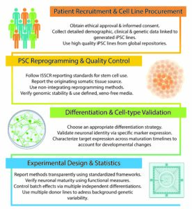
When recruiting donors for iPSC-based research projects, it is essential to clarify the need for ethical approval with local authorities and to provide donors with comprehensive information to obtain their informed consent. Information that should be provided in the consent materials has been suggested and summarized by Orzechowski et al. [44] and should include background information on the study, potential use of cells in future (commercial) research, storage of patient material and cell lines, as well as how to deal with incidental medical findings. Where possible, detailed demographic, clinical, medical, diagnostic, and genetic data should be collected and linked to the generated cell lines [13]. The chosen cohort for an iPSC-based study should be tailored to the specific research question of that study and strive for an appropriately representative, diverse sample.
In addition to reprogramming iPSC lines for a new study, already reprogrammed and quality-controlled iPSC lines from control or disease backgrounds can be obtained from various cell banks, repositories, or directly from the lab that generated and published the lines. Here is a list of global PSC banks: African iPSC Initiative, CellBank Australia, Coriell, European Bank for Induced Pluripotent Stem Cells, HipSci, Indian Stem Cell Repository, National Stem Cell Bank Korea, New York Stem Cell Foundation Repository, NINDS Human Cell and Data Repository, RIKEN BRC Japan, the Human Pluripotent Stem Cell Registry, UK Stem Cell Bank, WiCell.
Reprogramming and PSC quality control
Since the discovery of epigenetic reprogramming in 2006, iPSC technology has become a robust and widely adopted platform, supported by established protocols and quality control standards to ensure reproducibility, genomic integrity, and biological relevance. As such, several key considerations should be made when generating and using iPSC lines when modeling neuropathic pain mechanisms.
The source tissue used for reprogramming should be carefully selected and explicitly reported, as somatic cells may carry acquired genetic or epigenetic alterations that persist following reprogramming [43]. For example, dermal fibroblasts can potentially accumulate UV-associated or age-related mutations that may be retained in derived iPSC lines [40,56]. Where feasible, peripheral blood mononuclear cells (PBMCs) offer advantages, including ease of collection and reduced environmental mutational burden. Reprogramming should be performed, preferably using genome-non-integrating approaches, such as Sendai virus, episomal plasmids, or mRNA-based methods, to minimize insertional mutagenesis. iPSC lines should be assessed for genomic stability via karyotyping or copy-number analysis, and female donor–derived lines should be evaluated for X-chromosome inactivation status, especially when relevant to genetic pain studies [10]. Given the sensitivity of pluripotent stem cells to culture conditions and to ensure experimental reliability, iPSC cultures should be regularly screened for microbial contamination (e.g., mycoplasma), and the use of chemically defined, xeno-free media and matrices is required to reduce batch-to-batch variability and improve reproducibility across laboratories. The International Society for Stem Cell Research (ISSCR) recently published recommended standards for the use of human stem cells in research [55], as well as recommendations for reporting practices for publishing results with human pluripotent stem cells [47].
Differentiation strategies, quality control, and cell type validation
Multiple protocols have been published during the last 15 years for the in vitro derivation of human peripheral sensory neurons from pluripotent stem cells [27]. Most of these protocols can be divided into one of three approaches:
- Differentiation of sensory neurons from iPSCs guided by small molecules.
- Differentiation of sensory neurons from iPSCs guided by overexpression of fate-specifying transcription factors.
- Generation of a stable intermediate neural progenitor or neural crest cell population from iPSCs that can be further guided into sensory neurons or other neural crest derivatives.
Most published protocols generate a generic or mixed sensory neuron population [2,4,14,19,22,31,39]. However, some protocols report the generation of cultures enriched for nociceptive [3,7,11,46,51], mechanoreceptive [21,42,52] or proprioceptive [12,21] sensory neurons or to sort mixed sensory neuron cultures for specific subtypes after differentiation [49].
When implementing a differentiation protocol, validating and characterizing the generated cell type is very important. Identity of iSNs should be validated by testing for the co-expression of general sensory-neuron markers (e.g., peripherin, ISLET1, BRN3A) and subtype-specific markers (e.g., TRPV1, TRKA, Nav1.7, Nav1.8) [28]. In addition, electrophysiological functionality can be assessed by patch clamp and multielectrode array recordings in combination with specific pharmacological agonists or blockers [23,28]. A note of caution: stem-cell-derived in vitro models mimic human development; therefore, their state of maturation and functionality is a moving target that evolves during the course of culture. It has been shown that opioid receptors, while expressed early on in iSNs, initiate functional intracellular signaling only after prolonged culture times [48] and that robust membrane localization of Nav1.7 also only emerges over time in culture [32]. Therefore, when working with a novel target, it is advisable to characterize its expression, localization, and function in the chosen iSN model system over an extended period. Published transcriptome data of iSNs generated with existing, common protocols can be used to inform initial model selection [e.g., 33].
Statistical considerations and experimental design
As with in vivo research, translating findings from iPSC-based models to the clinic requires robust experimental design and appropriate statistical approaches, and several aspects must be considered. Primary outcomes should be defined a priori, and selected assays should directly measure the biological concepts under investigation [5,58]. Importantly, multiple complementary assays are often necessary to validate findings, especially during model development. Given the diversity of differentiation protocols used to generate sensory neurons, neuronal maturity must be carefully assessed and reported. Marker expression alone is insufficient to define functional maturity; therefore, ion channel function and excitability should also be evaluated, preferably using patch-clamp electrophysiology and/or multielectrode array (MEA) recordings, to ensure that developmental state does not introduce unintended variability [23]. Variability may also arise between differentiation rounds due to the multi-step nature of the process. To address this, data should be collected across multiple independent differentiations and several clones where feasible, and the differentiation batch should be considered during analysis.
Insights from other iPSC-based research fields indicate that genetic background is a major source of variability among reprogrammed iPSC lines [24,53]. This consideration is particularly important when modeling idiopathic or environmentally driven pain conditions, such as chemotherapy-induced peripheral neuropathy (CIPN). In such cases, experimental groups should include iPSC lines derived from multiple donors rather than relying on repeated measurements from a single line. Transcriptomic analyses suggest that groups often require four to six independent lines, although the appropriate sample size should be determined by model-specific power analysis [13,18,29]. In the case of monogenic pain disorders, such as Nav1.7 channelopathies, the use of isogenic controls provides an effective strategy to control for genetic background, provided that rigorous quality control is performed to exclude off-target effects of genome editing.
Beyond experimental design, accurate and consistent reporting of experimental parameters is essential for methodological rigor. Standardized reporting frameworks, such as the RIVER Recommendations [57], the FAIR guidelines [59], and the ENTRUST PE framework [16] provide practical guidance for transparent documentation and are strongly encouraged to improve the reproducibility of neuropathic pain models across laboratories.
Conclusion and Outlook
This fact sheet summarizes currently available methods and concepts for working with stem cell-derived models of neuropathic pain and aims to bridge pain researchers to resources available within the stem cell research community.
While iSN models have been optimized during the last decade and successfully employed to elucidate pathomechanisms and patient-specific phenotypes, some limitations and challenges remain: the lack of the natural niche of sensory neurons within our culture systems, the need for prolonged maturation times, and a reduced functionality in iSNs compared to primary hDRG neurons when it comes to certain pain targets, e.g., TRPV1 and Nav1.9. Optimized culture conditions and differentiation paradigms should help overcome these obstacles. In recent years, the number of more complex culture systems increased, including co-cultures of human iSNs with primary or iPSC-derived Schwann cells or satellite glial cells [4,8,22,30,54], iPSC-derived dorsal root ganglion organoids [34,35] and assembloids of multiple neuronal organoids reconstructing the somatosensory pathway [25]. Development and application of rigorous methods, as well as openly reporting and sharing differentiation and culture protocols within our research community, will help to further improve our model systems and research, bringing us closer to a better understanding of peripheral neuron development, function, and pathology.
Resource
https://youtu.be/iFfjQ5k7hqo?si=5b0y2wiiWNHGoV6g Watch an 8-minute video that walks through key resources, differentiation strategies, and practical tips for working with induced pluripotent stem cell (iPSC)-derived models in the context of neuropathic pain research, presented by Pascal Röderer, PhD.
References
- Alich TC, Röderer P, Szalontai B, Golcuk K, Tariq S, Peitz M, Brüstle O, Mody I. Bringing to light the physiological and pathological firing patterns of human induced pluripotent stem cell-derived neurons using optical recordings. Front Cell Neurosci 2023;16:676. doi:10.3389/FNCEL.2022.1039957.
- Alshawaf AJ, Viventi S, Qiu W, D’Abaco G, Nayagam B, Erlichster M, Chana G, Everall I, Ivanusic J, Skafidas E, Dottori M. Phenotypic and functional characterization of peripheral sensory neurons derived from human embryonic stem cells. Sci Rep 2018;8:603. doi:10.1038/s41598-017-19093-0.
- Boisvert EM, Engle SJ, Hallowell SE, Liu P, Wang Z-W, Li X-J. The specification and maturation of nociceptive neurons from human embryonic stem cells. Sci Rep 2015;5:16821. doi:10.1038/srep16821.
- Cai S, Han L, Ao Q, Chan Y-S, Shum DK-Y. Human Induced Pluripotent Cell-Derived Sensory Neurons for Fate Commitment of Bone Marrow-Derived Schwann Cells: Implications for Remyelination Therapy. Stem Cells Transl Med 2016;6:1–13. doi:10.5966/sctm.2015-0424.
- Cantor EL, Shen F, Jiang G, Philips S, Schneider BP. Optimization of a human induced pluripotent stem cell-derived sensory neuron model for the in vitro evaluation of taxane-induced neurotoxicity. Scientific Reports 2024 14:1 2024;14:19075-. doi:10.1038/s41598-024-69280-z.
- Cao L, McDonne A, Nitzsche A, Alexandrou A, Saintot PP, Loucif AJC, Brown AR, Young G, Mis M, Randal A, Waxman SG, Stanley P, Kirby S, Tarabar S, Gutteridge A, Butt R, McKernan RM, Whiting P, Ali Z, Bilsland J, Stevensl EB. Pharmacological reversal of a pain phenotype in iPSC-derived sensory neurons and patients with inherited erythromelalgia. Sci Transl Med 2016;8.
- Chambers SM, Qi Y, Mica Y, Lee G, Zhang XJ, Niu L, Bilsland J, Cao L, Stevens E, Whiting P, Shi SH, Studer L. Combined small-molecule inhibition accelerates developmental timing and converts human pluripotent stem cells into nociceptors. Nat Biotechnol 2012;30:715–720. doi:10.1038/nbt.2249.
- Clark AJ, Kaller MS, Galino J, Willison HJ, Rinaldi S, Bennett DLH. Co-cultures with stem cell-derived human sensory neurons reveal regulators of peripheral myelination. Brain 2017;140:898–913. doi:10.1093/brain/awx012.
- Clark AJ, Kugathasan U, Baskozos G, Priestman DA, Fugger N, Lone MA, Othman A, Chu KH, Blesneac I, Wilson ER, Laurà M, Kalmar B, Greensmith L, Hornemann T, Platt FM, Reilly MM, Bennett DL. An iPSC model of hereditary sensory neuropathy-1 reveals L-serine-responsive deficits in neuronal ganglioside composition and axoglial interactions. Cell Rep Med 2021;2:100345.
- DeBoever C, Li H, Jakubosky D, Benaglio P, Reyna J, Olson KM, Huang H, Biggs W, Sandoval E, D’Antonio M, Jepsen K, Matsui H, Arias A, Ren B, Nariai N, Smith EN, D’Antonio-Chronowska A, Farley EK, Frazer KA. Large-Scale Profiling Reveals the Influence of Genetic Variation on Gene Expression in Human Induced Pluripotent Stem Cells. Cell Stem Cell 2017;20:533-546.e7. doi:10.1016/j.stem.2017.03.009.
- Desiderio S, Vermeiren S, Van Campenhout C, Kricha S, Malki E, Richts S, Fletcher E V., Vanwelden T, Schmidt BZ, Henningfeld KA, Pieler T, Woods CG, Nagy V, Verfaillie C, Bellefroid EJ. Prdm12 directs nociceptive sensory neuron development by regulating the expression of the NGF Receptor TrkA. Cell Rep 2019;26:3522-3536.e5.
- Dionisi C, Rai M, Chazalon M, Schiffmann SN, Pandolfo M. Primary proprioceptive neurons from human induced pluripotent stem cells: a cell model for afferent ataxias. Sci Rep 2020;10:1–12.
- Dutan Polit L, Eidhof I, McNeill R V., Warre-Cornish KM, Yde Ohki CM, Walter NM, Sala C, Verpelli C, Radtke F, Galderisi S, Mucci A, Collo G, Edenhofer F, Castrén ML, Réthelyi JM, Ejlersen M, Hohmann SS, Ilieva MS, Lukjanska R, Matuleviciute R, Michel TM, de Vrij FMS, Kushner SA, Lendemeijer B, Kittel-Schneider S, Ziegler GC, Gruber-Schoffnegger D, Pasterkamp RJ, Kasri A, Potier MC, Knoblich JA, Brüstle O, Peitz M, Pich EM, Harwood AJ, Abranches E, Falk A, Vernon AC, Grünblatt E, Srivastava DP. Recommendations, guidelines, and best practice for the use of human induced pluripotent stem cells for neuropharmacological studies of neuropsychiatric disorders. Neuroscience Applied 2023;2:101125. doi:10.1016/J.NSA.2023.101125.
- Eberhardt E, Namer B, Neureiter A, Körner J, Jørum E, Kurth I, Winner B, Lampert A. Spontaneous activity in pain patient stem cell–derived sensory neurons arises from one functional subclass. Pain 2025. doi:10.1097/J.PAIN.0000000000003865.
- Engle SJ, Blaha L, Kleiman RJ. Best Practices for Translational Disease Modeling Using Human iPSC-Derived Neurons. Neuron 2018;100:783–797. doi:10.1016/J. NEURON.2018.10.033.
- ENTRUST-PE Framework. n.d. Available: https://entrust-pe.org/. Accessed 12 Feb 2026.
- Erbacher C, Andrews A, Sauerwein T, Breyer M, Arampatzi P, Koch M, Lamer S, Gräfenhan T, Schlosser A, Üçeyler N. Axon guidance deficits in a human sensory-like neuron model of Fabry disease. Exp Neurol 2026;397:115564. doi:10.1016/J. EXPNEUROL.2025.115564.
- Germain PL, Testa G. Taming Human Genetic Variability: Transcriptomic Meta-Analysis Guides the Experimental Design and Interpretation of iPSC-Based Disease Modeling. Stem Cell Reports 2017;8:1784–1796. doi:10.1016/J.STEMCR.2017.05.012.
- Guimarães MZP, De Vecchi R, Vitória G, Sochacki JK, Paulsen BS, Lima I, Rodrigues da Silva F, da Costa RFM, Castro NG, Breton L, Rehen SK. Generation of iPSC-Derived Human Peripheral Sensory Neurons Releasing Substance P Elicited by TRPV1 Agonists. Front Mol Neurosci 2018;11:277. doi:10.3389/fnmol.2018.00277.
- Hew L, Maierhof SK, Ivanov A, Beule D, Fernandez Vallone V, Stachelscheid H, Frahm S, Telugu NS, Endres M, Boehmerle W, Huehnchen P, Schinke C. c-Jun inhibition mitigates chemotherapy-induced neurotoxicity in iPSC-derived sensory neurons. Cell Death Discovery 2025 11:1 2025;11:529-. doi:10.1038/s41420-025-02847-5.
- Hulme AJ, Finol-Urdaneta RK, McArthur JR, Marzano NR, Maksour S, Thind A, Guo Y, Kaul D, Maddock M, Friedrich O, Martinac B, Adams DJ, Dottori M. Induced Proprioceptor and Low-Threshold Mechanoreceptor Neurons Derived from Human Pluripotent Stem Cells Exhibit Distinct Functional Mechanosensory Properties. Advanced Science 2025:e12413. doi:10.1002/ADVS.202512413;REQUESTEDJOURNAL:JOURNAL:21983844;WGROUP:STRING:PUBLICATION.
- Jones I, Yelhekar TD, Wiberg R, Kingham PJ, Johansson S, Wiberg M, Carlsson L. Development and validation of an in vitro model system to study peripheral sensory neuron development and injury. Sci Rep 2018;8:1–15. doi:10.1038/s41598-018- 34280-3.
- Kalia AK, Rösseler C, Granja-Vazquez R, Ahmad A, Pancrazio JJ, Neureiter A, Zhang M, Sauter D, Vetter I, Andersson A, Dussor G, Price TJ, Kolber BJ, Truong V, Walsh P, Lampert A. How to differentiate induced pluripotent stem cells into sensory neurons for disease modelling: a functional assessment. Stem Cell Research & Therapy 2024 15:1 2024;15:99-. doi:10.1186/S13287-024-03696-2.
- Kilpinen H, Goncalves A, Leha A, Afzal V, Alasoo K, Ashford S, Bala S, Bensaddek D, Casale FP, Culley OJ, Danecek P, Faulconbridge A, Harrison PW, Kathuria A, McCarthy D, McCarthy SA, Meleckyte R, Memari Y, Moens N, Soares F, Mann A, Streeter I, Agu CA, Alderton A, Nelson R, Harper S, Patel M, White A, Patel SR, Clarke L, Halai R, Kirton CM, Kolb-Kokocinski A, Beales P, Birney E, Danovi D, Lamond AI, Ouwehand WH, Vallier L, Watt FM, Durbin R, Stegle O, Gaffney DJ. Common genetic variation drives molecular heterogeneity in human iPSCs. Nature 2017 546:7658 2017;546:370–375. doi:10.1038/nature22403.
- Kim J, Imaizumi K, Jurjuț O, Kelley KW, Wang D, Thete MV, Hudacova Z, Amin ND, Levy RJ, Scherrer G, Pașca SP. Human assembloid model of the ascending neural sensory pathway. Nature 2025 2025:1–11. doi:10.1038/s41586-025-08808-3.
- Kolsters N, Vernon AC, Kasri NN, Latour BL. Establishing validity standards for iPSC modeling of neuropsychiatric disorders. Genomic Psychiatry 2025;1:27–23. doi:10.61373/GP025P.0074.
- Labau JIR, Andelic M, Faber CG, Waxman SG, Lauria G, Dib-Hajj SD. Recent advances for using human induced-pluripotent stem cells as pain-in-a-dish models of neuropathic pain. Exp Neurol 2022;358:114223.
- Lampert A, Bennett DL, McDermott LA, Neureiter A, Eberhardt E, Winner B, Zenke M. Human sensory neurons derived from pluripotent stem cells for disease modelling and personalized medicine. Neurobiology of Pain 2020:100055.
- Lazic SE, Clarke-Williams CJ, Munafò MR. What exactly is ‘N’ in cell culture and animal experiments? PLoS Biol 2018;16:e2005282. doi:10.1371/JOURNAL.PBIO.2005282.
- LeBlang CJ, Pazyra-Murphy MF, Silagi ES, Dasgupta S, Tsolias M, Miller T, Petrova V, Zhen S, Jovanovic VM, Castellano D, Gerrish K, Ormanoglu P, Tristan CA, Singeç I, Woolf CJ, Tasdemir-Yilmaz O, Segal RA. Satellite glial contact enhances differentiation and maturation of human iPSC-derived sensory neurons. Stem Cell Reports 2025;0:102639. doi:10.1016/J.STEMCR.2025.102639.
- Lee G, Chambers SM, Tomishima MJ, Studer L. Derivation of neural crest cells from human pluripotent stem cells. Nat Protoc 2010;5:688–701. doi:10.1038/ nprot.2010.35.
- Liu Y, Balaji R, de Toledo MAS, Ernst S, Hautvast P, Kesdoğan AB, Körner J, Zenke M, Neureiter A, Lampert A. The pain target NaV1.7 is expressed late during human iPS cell differentiation into sensory neurons as determined in high-resolution imaging. Pflugers Arch 2024;476:975–992. doi:10.1007/S00424-024-02945-W/FIGURES/7.
- Li Y, Lock A, Fedele L, Zebochin I, Sabate A, Siddle M, Cainarca S, Röderer P, Montag K, Tarroni P, Brüstle O, Shaw T, Taams L, Denk F. Modelling inflammation-induced peripheral sensitization in a dish—more complex than expected? Pain 2025. doi:10.1097/J.PAIN.0000000000003512.
- Lu T, Wang M, Zhou W, Ni Q, Yue Y, Wang W, Shi Y, Liu Z, Li C, Hong B, Zhou X, Zhong S, Wang K, Zeng B, Zhang J, Wang W, Zhang X, Wu Q, Wang X. Decoding transcriptional identity in developing human sensory neurons and organoid modeling. Cell 2024;187:7374-7393.e28. doi:10.1016/J.CELL.2024.10.023.
- Mazzara PG, Muggeo S, Luoni M, Massimino L, Zaghi M, Valverde PTT, Brusco S, Marzi MJ, Palma C, Colasante G, Iannielli A, Paulis M, Cordiglieri C, Giannelli SG, Podini P, Gellera C, Taroni F, Nicassio F, Rasponi M, Broccoli V. Frataxin gene editing rescues Friedreich’s ataxia pathology in dorsal root ganglia organoid-derived sensory neurons. Nat Commun 2020;11:1–18.
- McDermott LA, Weir GA, Themistocleous AC, Segerdahl AR, Blesneac I, Baskozos G, Clark AJ, Millar V, Peck LJ, Ebner D, Tracey I, Serra J, Bennett DL. Defining the functional role of NaV1.7 in human nociception. Neuron 2019;101:905-919.e8. doi:10.1016/J.NEURON.2019.01.047.
- Meents JE, Bressan E, Sontag S, Foerster A, Hautvast P, Rösseler C, Hampl M, Schüler H, Goetzke R, Chi Le TK, Kleggetveit IP, Le Cann K, Kerth C, Rush AM, Rogers M, Kohl Z, Schmelz M, Wagner W, Jørum E, Namer B, Winner B, Zenke M, Lampert A. The role of Nav1.7 in human nociceptors. Pain 2019.
- Mortensen C, Thomsen MT, Chua KC, Hammer HS, Nielsen F, Pötz O, Svenningsen AF, Kroetz DL, Stage TB. Modeling mechanisms of chemotherapy-induced peripheral neuropathy and chemotherapy transport using induced pluripotent stem cell-derived sensory neurons. Neuropharmacology 2024;258:110062. doi:10.1016/J. NEUROPHARM.2024.110062.
- Münst S, Koch P, Kesavan J, Alexander-Mays M, Münst B, Blaess S, Brüstle O. In vitro segregation and isolation of human pluripotent stem cell-derived neural crest cells. Methods 2018;133:65–80. doi:10.1016/J.YMETH.2017.09.012.
- Musunuru K, Sheikh F, Gupta RM, Houser SR, Maher KO, Milan DJ, Terzic A, Wu JC. Induced Pluripotent Stem Cells for Cardiovascular Disease Modeling and Precision Medicine: A Scientific Statement from the American Heart Association. Circ Genom Precis Med 2018;11:E000043. doi:10.1161/HCG.0000000000000043;PAGE:STRING :ARTICLE/CHAPTER.
- Namer B, Schmidt D, Eberhardt E, Maroni M, Dorfmeister E, Kleggetveit IP, Kaluza L, Meents J, Gerlach A, Lin Z, Winterpacht A, Dragicevic E, Kohl Z, Schüttler J, Kurth I, Warncke T, Jorum E, Winner B, Lampert A. Pain relief in a neuropathy patient by lacosamide: Proof of principle of clinical translation from patient-specific iPS cell-derived nociceptors. EBioMedicine 2019;39. Available: http://www.ncbi.nlm.nih. gov/pubmed/30503201. Accessed 28 Jan 2019.
- Nickolls AR, Lee MM, Espinoza DF, Szczot M, Lam RM, Wang Q, Beers J, Zou J, Nguyen MQ, Solinski HJ, AlJanahi AA, Johnson KR, Ward ME, Chesler AT, Bönnemann CG. Transcriptional programming of human mechanosensory neuron subtypes from pluripotent stem cells. Cell Rep 2020;30:932-946.e7. doi:10.1016/j. celrep.2019.12.062.
- Nishino K, Toyoda M, Yamazaki-Inoue M, Fukawatase Y, Chikazawa E, Sakaguchi H, Akutsu H, Umezawa A. DNA Methylation Dynamics in Human Induced Pluripotent Stem Cells over Time. PLoS Genet 2011;7:e1002085. doi:10.1371/JOURNAL. PGEN.1002085.
- Orzechowski M, Schochow M, Kühl M, Steger F. Donor information in research and drug evaluation with induced pluripotent stem cells (iPSCs). Stem Cell Res Ther 2020;11:126. doi:10.1186/S13287-020-01644-4.
- Perez-Sanchez J, Middleton SJ, Pattison LA, Hilton H, Ali Awadelkareem M, Zuberi SR, Renke MB, Hu H, Yang X, Clark AJ, St John Smith E, Bennett DL. A humanized chemogenetic system inhibits murine pain-related behavior and hyperactivity in human sensory neurons. Sci Transl Med 2023;15:eadh3839. doi:10.1126/SCITRANSLMED.ADH3839.
- Plumbly W, Patikas N, Field SF, Foskolou S, Metzakopian E. Derivation of nociceptive sensory neurons from hiPSCs with early patterning and temporally controlled NEUROG2 overexpression. Cell Reports Methods 2022;0:100341. doi:10.1016/J. CRMETH.2022.100341.
- Reporting Practices for Publishing Results with Human Pluripotent and Tissue Stem Cells. n.d. Available: https://www.isscr.org/basic-research-standards. Accessed 15 Jan 2026.
- Röderer P, Belu A, Heidrich L, Siobal M, Isensee J, Prolingheuer J, Janocha E, Valdor M, Hagendorf S, Bahrenberg G, Opitz T, Segschneider M, Haupt S, Nitzsche A, Brüstle O, Hucho T. Emergence of nociceptive functionality and opioid signaling in human induced pluripotent stem cell–derived sensory neurons. Pain 2023. Available: https://journals.lww.com/pain/Fulltext/9900/Emergence_of_nociceptive_functionality_and_opioid.249.aspx.
- Saito-Diaz K, Street JR, Ulrichs H, Zeltner N. Derivation of peripheral nociceptive, mechanoreceptive, and proprioceptive sensory neurons from the same culture of human pluripotent stem cells. Stem Cell Reports 2021;0. doi:10.1016/j.stemcr.2021.01.001.
- Schinke C, Vallone VF, Ivanov A, Peng Y, Körtvelyessy P, Nolte L, Huehnchen P, Beule D, Stachelscheid H, Boehmerle W, Endres M. Modeling chemotherapy induced neurotoxicity with human induced pluripotent stem cell (iPSC) -derived sensory neurons. Neurobiol Dis 2021;155:105391.
- Schrenk-Siemens K, Pohle J, Rostock C, Abd El Hay M, Lam RM, Szczot M, Lu S, Chesler AT, Siemens J. Human Stem Cell-Derived TRPV1-Positive Sensory Neurons: A New Tool to Study Mechanisms of Sensitization. Cells 2022, Vol 11, Page 2905 2022;11:2905. doi:10.3390/CELLS11182905.
- Schrenk-Siemens K, Wende H, Prato V, Song K, Rostock C, Loewer A, Utikal J, Lewin GR, Lechner SG, Siemens J. PIEZO2 is required for mechanotransduction in human stem cell–derived touch receptors. Nat Neurosci 2015;18:10–16. doi:10.1038/ nn.3894.
- Schwartzentruber J, Foskolou S, Kilpinen H, Rodrigues J, Alasoo K, Knights AJ, Patel M, Goncalves A, Ferreira R, Benn CL, Wilbrey A, Bictash M, Impey E, Cao L, Lainez S, Loucif AJ, Whiting PJ, Gutteridge A, Gaffney DJ, HIPSCI Consortium DJ. Molecular and functional variation in iPSC-derived sensory neurons. 2018;50:54–61. doi:10.1038/s41588-017-0005-8.
- Shi L, Huang L, He R, Huang W, Wang H, Lai X, Zou Z, Sun J, Ke Q, Zheng M, Lu X, Pei Z, Su H, Xiang AP, Li W, Yao X. Modeling the Pathogenesis of Charcot-Marie-Tooth Disease Type 1A Using Patient-Specific iPSCs. Stem Cell Reports 2018;10:120–133.
- Standards for Human Stem Cell Use in Research. n.d. Available: https://www.isscr. org/basic-research-standards. Accessed 15 Jan 2026.
- Strässler ET, Aalto-Setälä K, Kiamehr M, Landmesser U, Kränkel N. Age Is Relative— Impact of Donor Age on Induced Pluripotent Stem Cell-Derived Cell Functionality. Front Cardiovasc Med 2018;5:325922. doi:10.3389/FCVM.2018.00004/BIBTEX.
- The RIVER working group. Reporting In Vitro Experiments Responsibly – the RIVER Recommendations. 2023. doi:10.31222/OSF.IO/X6AUT.
- Volpato V, Webber C. Addressing variability in iPSC-derived models of human disease: Guidelines to promote reproducibility. DMM Disease Models and Mechanisms 2020;13. doi:10.1242/DMM.042317/223104.
- Wilkinson MD, Dumontier M, Aalbersberg IjJ, Appleton G, Axton M, Baak A, Blomberg N, Boiten JW, da Silva Santos LB, Bourne PE, Bouwman J, Brookes AJ, Clark T, Crosas M, Dillo I, Dumon O, Edmunds S, Evelo CT, Finkers R, Gonzalez-Beltran A, Gray AJG, Groth P, Goble C, Grethe JS, Heringa J, t Hoen PAC, Hooft R, Kuhn T, Kok R, Kok J, Lusher SJ, Martone ME, Mons A, Packer AL, Persson B, Rocca-Serra P, Roos M, van Schaik R, Sansone SA, Schultes E, Sengstag T, Slater T, Strawn G, Swertz MA, Thompson M, Van Der Lei J, Van Mulligen E, Velterop J, Waagmeester A, Wittenburg P, Wolstencroft K, Zhao J, Mons B. The FAIR Guiding Principles for scientific data management and stewardship. Scientific Data 2016 3:1 2016;3:160018-. doi:10.1038/sdata.2016.18.
- Yang NJ, Isensee J, Neel D V., Quadros AU, Zhang H-XB, Lauzadis J, Liu SM, Shiers S, Belu A, Palan S, Marlin S, Maignel J, Kennedy-Curran A, Tong VS, Moayeri M, Röderer P, Nitzsche A, Lu M, Pentelute BL, Brüstle O, Tripathi V, Foster KA, Price TJ, Collier RJ, Leppla SH, Puopolo M, Bean BP, Cunha TM, Hucho T, Chiu IM. Anthrax toxins regulate pain signaling and can deliver molecular cargoes into ANTXR2+ DRG sensory neurons. Nature Neuroscience 2022 2021:1–12. doi:10.1038/s41593-021- 00973-8.
Disclosures
ALa receives councelling fees from Grünenthal and Orion.