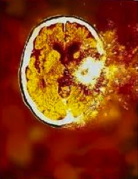
Perhaps our language has always hinted at the involvement of glial cells in injury? And, when glial cells outnumber neurons in the brain by 20 to 1, it begs the question who is really in charge of synaptic activity (should that be plasticity) in the brain?
I think it is fair to say that ever since the neuronal doctrine captured the imagination of neuroscientists at the end of the 1880s, when Ramon y Cajal made the seminal statement that “each nerve cell is a totally autonomous physiological canton”(Verkhratsky, Parpura et al. 2010), we have approached synaptic plasticity from a neurocentric perspective. In times of synaptic change such as injuring forces and other more commonplace examples such as memory and learning we have developed models of neural circuits, neuronal and molecular change to explain how the change occurs. However, there is now a large body of evidence we need to embrace to ensure we don’t continue to dismiss glial involvement in these changes. They are not a bunch of redundant polystyrene-like packing baubles forming fluffy sofas for all the important cells to recline on.
At the recent NOI conference Mick Thacker waxed lyrical about glia. One concept he developed with us, and that I am going to explore here, was that of considering the synaptic cleft in the brain as a three way interaction: the pre and post synaptic surfaces of the neurons and the astrocyte that envelops the synapse (for more on this have a look at (Araque, Parpura et al. 1999; Verkhratsky, Parpura et al. 2010). The astrocyte is not merely a decorative scarf – it can change the flavour of the chemical soup within the synapse by listening and talking to neurons and has continuous involvement synaptic transmission. To quote Verhratsky et al (2010) “Removal of the astroglial coverage results in synaptic shutdown and synapse elimination.” So who is in control of synaptic plasticity?
Some of the functions we know the astrocyte to be capable of: it can listen and talk to all the chemical messages in the central neural network because it is able to express virtually every receptor to neuroproteins – neurotransmitters, neurohumoral factors and neuromodulators (different combinations are expressed depending on use in different regions of the brain); it has a leading role to maintain extracellular concentrations of destabilising potassium (K+) around the synapse (increased by frequent neural firing and maybe too many bananas?) – it takes it up and breaks K+ down or redistributes it to areas of low K+; it collects glutamate, turns it into inert glutamine and transports it back to the extracellular space, ready for the neuron to resorb it and remake it into glutamate for future use; it gets excited when a majority of its receptors are activated by spill over of excitatory chemicals in the synapse and activates microglia (who fluff up and move around the brain to let us know something is going on); astrocytes contribute to the excitatory milieu with a chemical release of their own – cytokines and chemokines; astrocytes bridge the neuron/blood vessel interaction by having a foot in each camp literally and regulating local blood flow depending on the soup flavour at the time; they provide local metabolic and trophic support to neurons and in particular release TNF if there has been prolonged inactivity in the synapse – causing dendritic branching and new synapse formation via a variety of mechanisms (McAfoose and Baune 2009; Verkhratsky, Parpura et al. 2010).
Synaptic transmission is governed by the volume of chemicals in the synaptic space and the astrocytes and microglia govern this, so who is in charge? Clearly it is time to give deference to the glial cells, expand our neurocentric perspective and start considering the impact neuroglial cells make on the clinical stories we hear. The upregulation of central synaptic activity at the time of perceived or actual threat or injury, the formation of memory or acts of learning, that serve to make us aware of our surroundings and take action can now be interpreted in the light of not only neural excitability, but glial cell activation as well.
To be continued…
Carolyn Berryman
 Carolyn has been teaching with the Noisters (Neuro Orthopaedic Institute) for the last 10 years and finally got herself into research via a very competitive post-graduate scholarship. Not that she has ever stopped studying – she already has a masters in physiotherapy and in pain science – good luck fitting PhD onto the business card! What is Carolyn researching for her PhD? Based at the University of South Australia in Adelaide Carolyn is looking at neurophysiological profiles between chronic pain and PTSD (post traumatic stress disorder) during working memory tasks.
What does Carolyn do day to day? She is under piles of papers doing a systematic review of working memory and cognitive impairment in chronic pain. Carolyn will then be using EEG to evaluate what happens in people with pain. When she is not in the office, she lives on an island 100 kms south of Adelaide (that’s a long commute!) and spends her off-time playing with family, sailing and walking. Here is Carolyn talking more about the research she is doing.
References
Ader R, & Cohen N (1975). Behaviorally conditioned immunosuppression. Psychosomatic medicine, 37 (4), 333-40 PMID: 1162023
Araque A, Parpura V, Sanzgiri RP, & Haydon PG (1999). Tripartite synapses: glia, the unacknowledged partner. Trends in neurosciences, 22 (5), 208-15 PMID: 10322493.
Hayden, J., M. van Tulder, et al. (2011). “Exercise therapy for treatment of non-specific low back pain.” The Cochrane Library(2).
Hong, S. (2011). Can we jog our way to a younger-looking immune system? Brain, Behavior, and Immunity, 25 (8), 1519-1520 DOI: 10.1016/j.bbi.2011.08.002
McAfoose, J., & Baune, B. (2009). Evidence for a cytokine model of cognitive function Neuroscience & Biobehavioral Reviews, 33 (3), 355-366 DOI: 10.1016/j.neubiorev.2008.10.005
Verkhratsky, A., Parpura, V., & Rodríguez, J. (2011). Where the thoughts dwell: The physiology of neuronal–glial “diffuse neural net” Brain Research Reviews, 66 (1-2), 133-151 DOI: 10.1016/j.brainresrev.2010.05.002


