Editor’s note: The following is a summary of the John Bonica Named Lecture delivered at the Australian Pain Society 39th Annual Scientific Meeting. John Bonica—widely regarded as the founding father of pain medicine—strove to provide an egalitarian, interdisciplinary, and international forum to improve all aspects of pain, from research and treatment to education. This passion led to the incorporation of the International Association for the Study of Pain (IASP) in May 1974. The Bonica Lecture has been a feature of the Australian Pain Society Annual Scientific Meeting since Bonica himself gave the first lecture in 1984. The 2019 Bonica Lecture was delivered by Glenn King, a biochemist and structural biologist from the University of Queensland, at the recent meeting, which took place April 7 to 10 on the Gold Coast, Queensland, Australia.
Animals can use venom to capture their prey, but spiders and other venomous creatures also use their venom to deter predators. Inflicting pain via a venom is a highly effective defense mechanism that teaches potential predators (and their offspring) to stay away. This is like accidentally touching a hot stove—you live to tell the tale, don’t do it again, and can warn others of the danger.
But does what we see occurring in nature apply to the context of chronic pain in people? This was the focus of the John Bonica Named Lecture at the 2019 Annual Scientific Meeting of the Australian Pain Society, which was delivered by Glenn King, University of Queensland. King made the argument that animal venoms could potentially be used to understand pain perception in people and serve as a source of safer and more effective pain relievers.
Anatomically simple, molecularly complex
King started by describing how the anatomical pathway involved in nociceptive signaling is reasonably simple to understand. Peripheral nociceptors detect noxious stimuli and transmit this information to the dorsal horn of the spinal cord. The information is then sent up to the brain, where it may or may not be perceived as painful depending on both the prior and current context surrounding a potentially painful event. The brain then uses either descending facilitation or inhibition to elicit a response from the organism.
But, according to King, the molecular circuitry and signaling involved in nociception are far more complex in comparison, with a vast range of receptors and ion channels implicated to date.
“Trying to interpret all of this is like trying to figure out what is going on in a circuit board when you don’t know what all of the components are,” King explained. “Many of the ion channels and receptors involved in nociceptive processes have only been identified in the last 10 to 15 years, and there’s clearly more stuff that we don’t know about.” King is interested in whether animal venoms can help us complete the circuit board—by identifying more of the components—and enhance our understanding of nociceptive processing.
How venoms interact with pain pathways
To show how venoms affect nociceptive pathways, King and his team take dorsal root ganglia (DRG) neurons from mice, plate the cells, and then add a fluorescent calcium indicator, which serves as a measure of neuronal activity.
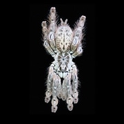
According to this assay, venom from some animals such as the bullet ant, Paraponera clavata, can robustly stimulate all sensory neurons within three seconds. However, the venom from other animals like the Togo starburst tarantula, Heteroscodra maculata, activates most, but not all sensory neurons. Animals with this kind of venom are of great interest to researchers like King.
“If you activate just a subset of the neurons it is much more likely you are activating a specific receptor or receptors, rather than activating every neuron,” King said. Indeed, this will make it easier to identify particular ion channels and other molecules that venoms may work through to produce pain.
Through collaboration with David Julius, University of California, San Francisco, US, King and his team identified two homologous peptides from the venom of the Togo starburst tarantula, Hm1a and Hm1b, as capable of activating DRG neurons (Osteen et al., 2016). These peptides are incredibly stable due to their unique 3D protein structure containing three disulfide bonds (Richards et al., 2018). This means that Hm1a and Hm1b are not easily broken down in blood or gastric fluids, and are highly resistant to heat and human chemicals.
Interestingly, neuronal activation by Hm1a is completely blocked by tetrodotoxin, a highly potent toxin that binds to voltage-gated sodium channels and is found in a range of animals such as fugu (Japanese puffer fish) and blue-ringed octopuses. The antagonism of Hm1a activity by tetrodotoxin suggests that Hm1a acts on those sodium channels, which play an important role in the initiation of action potentials in sensory neurons, including primary afferent pain fibers.
You can’t judge a book by its cover
“We initially thought, that’s really boring—it’s just another sodium channel toxin,” King recalled. Humans have nine different voltage-gated sodium (Nav) channels found in the heart, muscles, and the central and peripheral nervous system. Three of these channels, Nav1.7, Nav1.8, and Nav1.9, are well known for their contribution to nociceptive signaling. In addition, many commonly used analgesics such as amitriptyline and lidocaine are non-selective Nav blockers. This explains the side effects of many of these medications, as they end up acting on Nav channels in many different physiological systems.
“We believed that Hm1a was most likely activating Nav1.7 channels, because of the known gain-of-function mutations in this receptor leading to episodic pain disorders,” King said. However, it turned out that Hm1a was a highly selective activator of the Nav1.1 channel, keeping the channel open. Nav1.1 had not previously been implicated in pain processing. It is located primarily in medium-diameter DRG neurons, most likely myelinated Aδ fibers, where it is highly co-localized with Nav1.7.
King and his team then looked at c-Fos expression, a surrogate marker of neuronal activity, in the spinal cord of mice. Interestingly, the peptide led to neuronal activation in the spinal cord only on the side where the researchers had injected the peptide. But Hm1a elicited severe bilateral mechanical hypersensitivity, according to mechanical threshold testing. There was no effect of Hm1a on heat or inflammatory pathways.
“This is really cool. These results suggest that the Nav1.1 channel is involved in the transduction of mechanical stimuli to the spinal cord,” said King.
A mouse model of IBS
In collaboration with Stuart Brierley, South Australian Health and Medical Research Institute, Adelaide, who has developed a mouse model of irritable bowel syndrome (IBS), King then asked whether Nav1.1 is involved in mechanical pain in the gut during IBS, which is a common feature of the condition. Using Brierley’s model, the researchers saw upregulation of Nav1.1 channel expression in colonic DRG neurons in IBS mice, with up to 50 percent of neurons expressing it.
“The obvious clinical question then became whether or not a Nav1.1 channel inhibitor could block the mechanical gut pain associated with IBS,” said King.
So King and his colleagues returned to animal venoms in search of such an inhibitor. After setting up an assay to compare tens of thousands of peptides from different animal venoms, they identified the spider venom peptide Hs1a as an equipotent inhibitor of both the Nav1.1 and Nav1.7 channels (Klint et al., 2015).
In a separate assay of spider venoms performed by Irina Vetter, University of Queensland, and her team, a molecule known as Pn3a was identified as a highly specific inhibitor of the Nav1.7 channel but not the Nav1.1 channel (Deuis et al., 2017). King, Brierley, and colleagues could now use the Hs1a and Pn3a peptides to examine how the Nav1.1 and Nav1.7 channels contributed to mechanical pain in the IBS mouse model.
When the researchers tested the effects of Hs1a, Pn3a, and ICA-121431 (a selective Nav1.1 channel blocker), they saw that Hs1a, by blocking both Nav1.1 and Nav1.7, was more effective than Pn3a or ICA-121431 at reducing mechanical hypersensitivity in the IBS mouse model (unpublished findings). Hs1a was as effective as lidocaine in reversing the colonic hypersensitivity seen in the IBS mouse model but had the added benefit of minimal impact on colonic motility, a common side effect of lidocaine. King believes that peptides such as Hs1a would work effectively if taken orally as a tablet, but further testing of Hs1a is required before it can be considered for use in humans.
In the end, it’s clear that venoms from spiders and other creatures have much to teach us about pain processing, both in animals and in people. And as the work with the dual Nav1.1/Nav1.7 channel blocker Hs1a shows, they also point the way to potential novel analgesic treatments.
Lincoln Tracy is a postdoctoral research fellow in the School of Public Health and Preventive Medicine at Monash University and a freelance writer from Melbourne, Australia. You can follow him on Twitter @lincolntracy.
Image: Heteroscodra maculata (Togo starburst tarantula). Credit: ArachnoServer Spider Toxin Database/Attribution-NonCommercial 3.0 Unported (CC BY-NC 3.0).


